need help with your account or subscription? click here to email us (or see the contact page)
join telegramNEW! discord
jump to exam page:
search for anything ⋅ score predictor (โ€œpredict me!โ€)

NBME 22 Answers

nbme22/Block 1/Question#37 (reveal difficulty score)
A 28-year-old woman is brought to the ...
Area labeled โ€˜Cโ€™ (Abducens nucleus, right) ๐Ÿ” / ๐Ÿ“บ / ๐ŸŒณ / ๐Ÿ“–
tags:

 Login (or register) to see more


 +29  upvote downvote
submitted by โˆ—imgdoc(183)
get full access to all contentpick a username

I think its the right MLF (area C), not the right abducens nucleus that is lesioned on the cross section. If the abducens nucleus were lesioned she wouldn't have abduction in her left eye. The MLF mediates cross talk between the abducens nucleus on both sides to the MLF on the opposite side (2 abducens nuclei, 2 MLF one on each side). In her case, her right MLF wasn't functioning hence why she was gazing left but her right middle rectus wasn't contacting to mediate leftward gaze.

This picture helps: http://what-when-how.com/wp-content/uploads/2012/04/tmp15F9_thumb.jpg

get full access to all contentpick a username
imgdoc  *medial rectus +1

Quick review of gaze control; using leftward gaze as example

  • horizontal gaze control stems from left frontal gaze center
  • signal crosses to right pontine gaze center
  • signal goes to right MLF that yokes together right eye CN III to adduct and left eye CN VI to abduct and look to the left

Correction to myself sorry, wish could edit/delete I got the crossing mixed

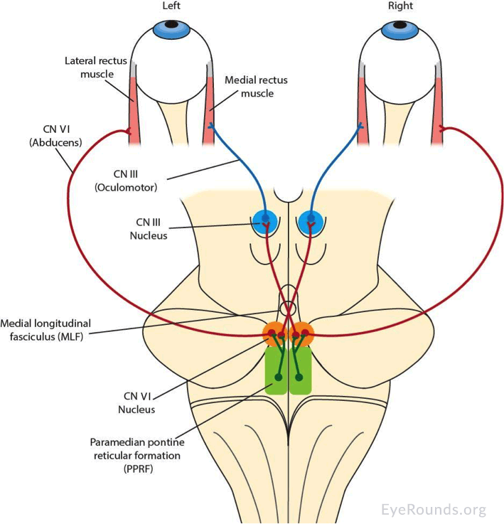
For left gaze + horizontal gaze stems from RIGHT frontal gaze center + crosses to LEFT pontine gaze center + left pontine gaze center links to left CN VI to abduct left eye; sends crossing to link with the RIGHT MLF + RIGHT MLF then adducts the right eye via CN III


 +7  upvote downvote
submitted by โˆ—lnsetick(107)
get full access to all contentpick a username

This is internuclear ophthalmoplegia. In INO you would see impaired adduction during horizontal gaze due to a lesion of the ipsilateral MLF. However, since the MLF is not involved in convergence, the affected eye is still able to converge normally.

get full access to all contentpick a username


 +6  upvote downvote
submitted by ahd_ve(7)
get full access to all contentpick a username

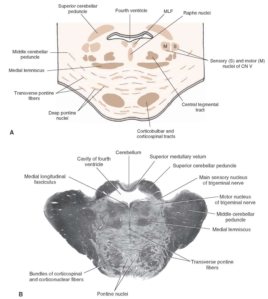
This imagen is a transverse section through the pons at the level of trigeminal nuclei: A and B: Superior Cerebellar peduncle. C and D: Medial longinutidinal fasciculus. E and F: Bundles of corticospinal and cortinuclear fibers.

get full access to all contentpick a username


 +4  upvote downvote
submitted by โˆ—drdoom(1206)
get full access to all contentpick a username

Hereโ€™s another very nice one that superimposes the pathway onto a simplified brainstem drawing (nice for the anatomical relations):

https://webeye.ophth.uiowa.edu/eyeforum/cases-i/case252/Fig2-INO-LRG.png

Source article:

https://webeye.ophth.uiowa.edu/eyeforum/cases/252-internuclear-ophthalmoplegia.htm

To see even more, try google image search on โ€œmedial longitudinal fasciculusโ€:

https://www.google.com/search?q=medial+longitudinal+fasciculus&tbm=isch

get full access to all contentpick a username
endochondral1  what is A and B in this pic? i knew it was dorsal pons ipsilateral but i just didn't know what part that was on the pic? +1
nwinkelmann  A and B are the superior cerebellar peduncles.http://what-when-how.com/wp-content/uploads/2012/04/tmp15F2.jpg +1


 +4  upvote downvote
submitted by โˆ—hyperfukus(111)
get full access to all contentpick a username

so the lesion is in the Right MLF right? If so I'm just about to memorize the eye see SAME MiLF lol its the MLF on the same side of the eye keep it simple i hope that's what yall are saying lol

get full access to all contentpick a username


 +1  upvote downvote
submitted by โˆ—drdoom(1206)
get full access to all contentpick a username

Nice schematic of how horizontal gaze is coordinated through the abducens/MLF/oculomotor pathway:

https://n.neurology.org/content/neurology/70/17/e57/F1.large.jpg

In the diagram, the system is coordinating gaze toward ptโ€™s left, which (conveniently) is the same as in the stem.

Source article: https://n.neurology.org/content/70/17/e57

get full access to all contentpick a username


 +0  upvote downvote
submitted by โˆ—pppro(25)
get full access to all contentpick a username

The lesion is in the medial longitudinal fasiculus.

http://what-when-how.com/neuroscience/brainstem-iii-the-midbrain-organization-of-the-central-nervous-system-part-1/

get full access to all contentpick a username


 +0  upvote downvote
submitted by โˆ—hello(429)
get full access to all contentpick a username

If you need a different orientation for a diagram of the pathways:

https://media.springernature.com/full/springer-static/image/art%3A10.1038%2Fs41433-018-0216-y/MediaObjects/41433_2018_216_Fig1_HTML.png?as=webp

get full access to all contentpick a username


 +0  upvote downvote
submitted by โˆ—okokok1(38)
get full access to all contentpick a username

The question stem is describing a pt with Multiple Sclerosis. INO is HIGHLY associated with that. Then if you follow the rule of 4's, you know that the MLF is medial and ipsilateral to the affected side. Thus, it must be the medial right structure.

get full access to all contentpick a username


Must-See Comments from nbme22

sacredazn on Unrearranged immunoglobulin gene
seagull on Decreased binding of RNA polymerase
seagull on Anticholinergic
liverdietrying on Release of stored thyroid hormone from a ...
keycompany on Negative nitrogen balance
kernicterusthefrog on Displacement
mcl on Area labeled โ€˜Dโ€™
joha961 on Displacement
imgdoc on Area labeled โ€˜Cโ€™ (Abducens nucleus, right)
alwaysanonymous on 25 mL/cm H2O
drdoom on 1 in 600
seagull on Glutamine
bubbles on Acute retroviral infection
yotsubato on Phase variation

search for anything NEW!