need help with your account or subscription? click here to email us (or see the contact page)
join telegramNEW! discord
jump to exam page:
search for anything ⋅ score predictor (โ€œpredict me!โ€)

NBME 16 Answers

nbme16/Block 1/Question#41 (reveal difficulty score)
An 18-year-old man with acute lymphoblastic ...
Amphotericin B ๐Ÿ” / ๐Ÿ“บ / ๐ŸŒณ / ๐Ÿ“–
tags:

 Login (or register) to see more


 +4  upvote downvote
submitted by โˆ—dentist(94)
get full access to all contentpick a username

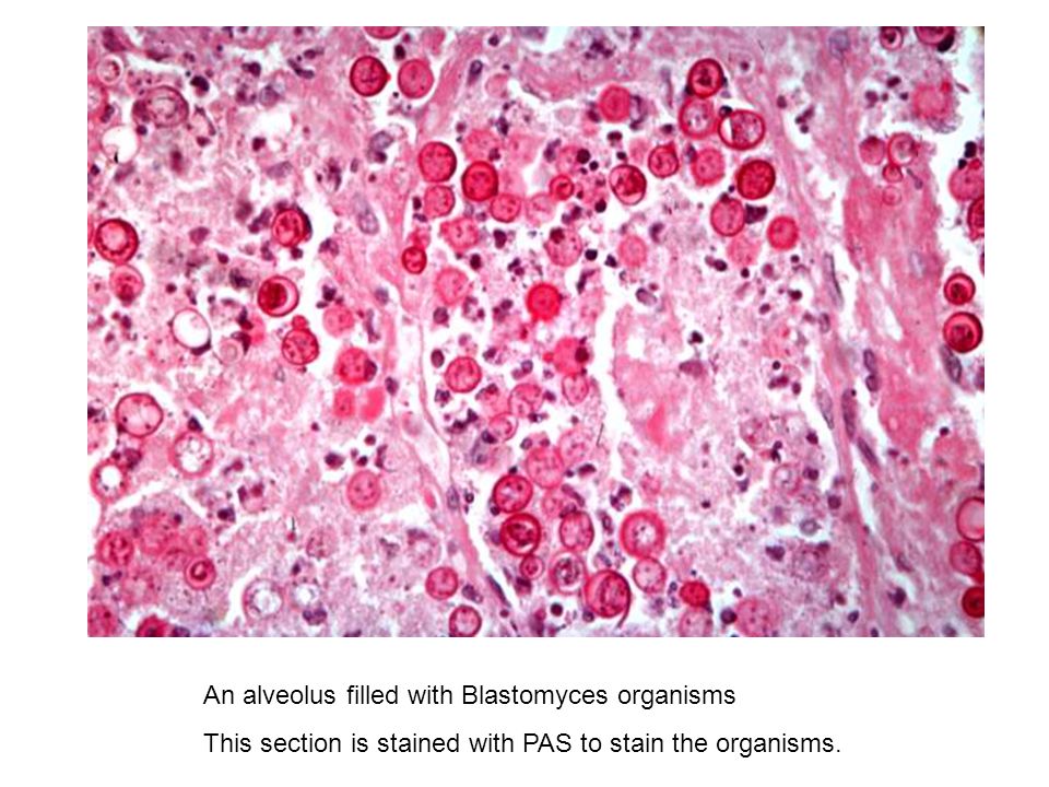
The solid red bits are of interest here: We're looking at blasto yeast buds (yeast at 37C). Always compare to RBC:

Blasto = RBC size

Coccidio > RBC

Histo < RBC

"history < of blasting (rbc) < coc(c)k" dont @ me

get full access to all contentpick a username


 +2  upvote downvote
submitted by โˆ—lsp1992(24)
get full access to all contentpick a username

Can someone please explain what we're seeing on the histo slide? I chose the correct answer because I was thinking fungus because of the immunocompromise and neutropenia (and I thought PAS was used for aspergillus), but I don't see anything fungus-related on that slide.

get full access to all contentpick a username
cassdawg  I think this is Blastomyces (broad based buds) where the darker pink are the blastomyces budding. Here are some similar slides: https://images.slideplayer.com/25/7691707/slides/slide_39.jpg https://www.gettyimages.co.uk/detail/photo/blastomycosis-in-the-brain-caused-by-the-high-res-stock-photography/vis303384 It could also be cryptococcus potentially (https://www.omicsonline.org/publication-images/diagnostic-pathology-budding-cryptococci-3-139-g005.png) but I think the bud bases are too broad and there is no clearing/visible capsule that cryptococcus is notable for. Either way you treat both systemic mycoses with amphotericin. +10
passplease  How did you eliminate CMV? +2
cassdawg  For me, CMV would have the characteristic "owl eye intranuclear inclusion" cells on biopsy but would be less likely to show anything in pleural fluid (i.e. thoracocentesis would not be used to diagnose CMV). Further CMV pneumonitis is an atypical/interstitial pneumonitis (diffuse patchy infiltrates on CXR, FA2020 p683) and he has a lower lobe consolidation with pleural effusion (more characteristic of fungal pneumonia). +6
shervinbd  Looks like Cryptococcus neoformans to me. +4
chaosawaits  I was pretty confident that this is Cryptococcus but the more I look at it, the more I don't know +1


Must-See Comments from nbme16

zincy7 on Gastrin
bingcentipede on Cerebral edema
cassdawg on Kidney
medninja on Lysosomes
cassdawg on Sildenafil
cassdawg on Feces-contaminated soil
cassdawg on Peutz-Jeghers
cassdawg on Presence of an internal ribosome entry site
hungrybox on Induction of cytochrome P450 enzymes that ...
andro on Antagonizes VLDL-cholesterol secretion
bingcentipede on Decreased function of Na+โ€“K+ ATPase
bingcentipede on Glyceraldehyde-3-phosphate dehydrogenase
cassdawg on 46,XXY
bingcentipede on Grade 4/6, holosystolic murmur heard best ...

search for anything NEW!